How a Proton Treatment Device Works
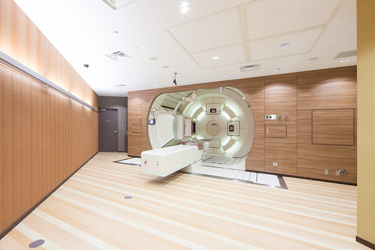
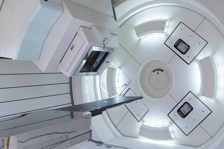
Gantry Irradiation Chambers 1 & 2
These irradiation chambers can focus the proton beams anywhere by rotating the gantries 360 degrees. It is possible to avoid areas we do not wish to irradiate and project the beams at the back or the sides of the body, or any other angle desired.
Gantry Irradiation Chamber 1 features the first spot scanning irradiation in Japan. This irradiation method is suited for treating the head and neck, pelvis, spine, and other areas.
Gantry Irradiation Chamber 2 features a double-scattering irradiation device. In this chamber, treatment is mainly performed on the lungs or liver.
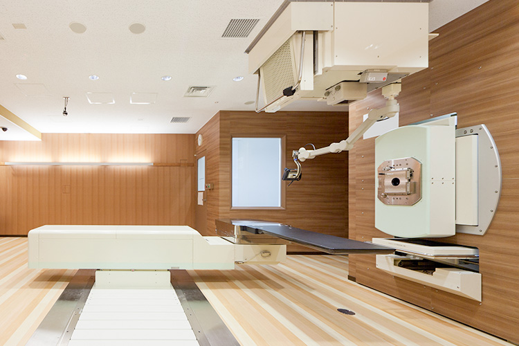
Fixed Irradiation Chamber
The fixed irradiation chamber features a double-scattering irradiation device. This chamber allows proton beams to be irradiated horizontally, and is mainly used in treating prostate cancer.
What is “spot scanning irradiation”?
Spot scanning irradiation is an advanced method that uses narrow proton beams sent from the accelerator to bathe the cancerous areas in radiation. This way, the irradiation can be matched to the shape of the cancer, and it is also possible to reduce damage to normal tissue.
What is “double-scattering irradiation”?
By scattering narrow proton beams sent from the accelerator, the protons are spread out over an area 14-25 cm in diameter. After that, by using a multi-leaf collimator or a carved collimator assembled for each patient, the proton beam is shaped to border the cancer. In the depth direction, a bolus is created for each patient and reproduces the shape of the cancer. This allows us to fit the proton beam in three dimensions to the shape of the cancer, and reduce the effects on healthy tissue during irradiation.
Multi-leaf collimator
Dozens of thin steel blades controlled by computer are adjusted for each patient, matching the irradiating proton beam to the shape of the cancer.
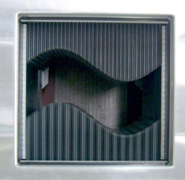
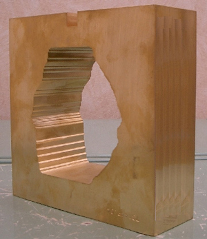
Carved collimator
This is carved from a block of brass using a special machine. Each one is constructed individually for each patient.
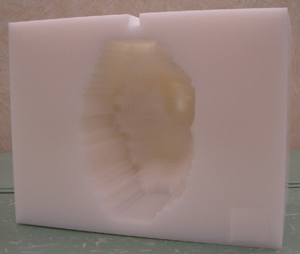
Bolus
This is carved out of a block of polyethylene using a special machine. The depth is matched to the shape of the cancer.
Determining positioning in the irradiation chamber
X-ray images can be used to control the position of the body with an accuracy of within 0.5 mm. By positioning the body accurately, it is possible to accurately aim the protons at the cancer and achieve the most precise treatment.
Gantry
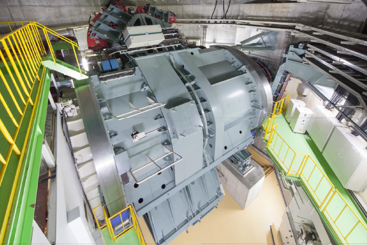
This massive machine, with an internal diameter of 5 m and a weight of 200 tonnes, is where the proton beams are bent. By rotating this machine, we can aim protons at the tumor from a full 360 degrees of movement, which means we can select the direction that will cause the least harm to the healthy tissue. By rotating it with an extremely high degree of accuracy, we can hit spheres as small as 2 mm across.
Injector (Ion Source, Linac)
The injector uses two different devices, an ion source and a linear particle accelerator (linac), to accelerate proton beams to 10% of the speed of light. We use hydrogen gas as our raw material, creating protons using electricity and then accelerating them.
Hydrogen is made up of a proton and an electron circling it. In the ion source, we use electrical discharges to turn the hydrogen into plasma, and extract the protons.
The extracted protons carry a positive electrical charge, so we can accelerate them using electrical force (Coulomb force). In the linac we use high-frequency electrical fields to accelerate protons about 5 m in a direct line.
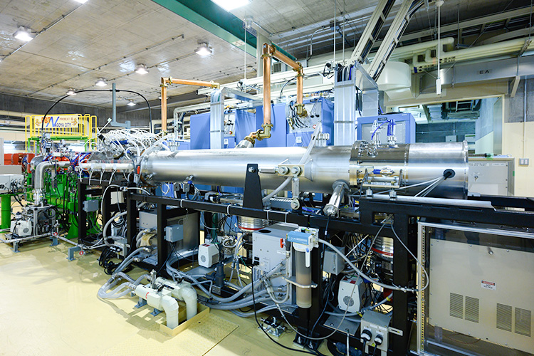
Accelerator (Synchrotron)
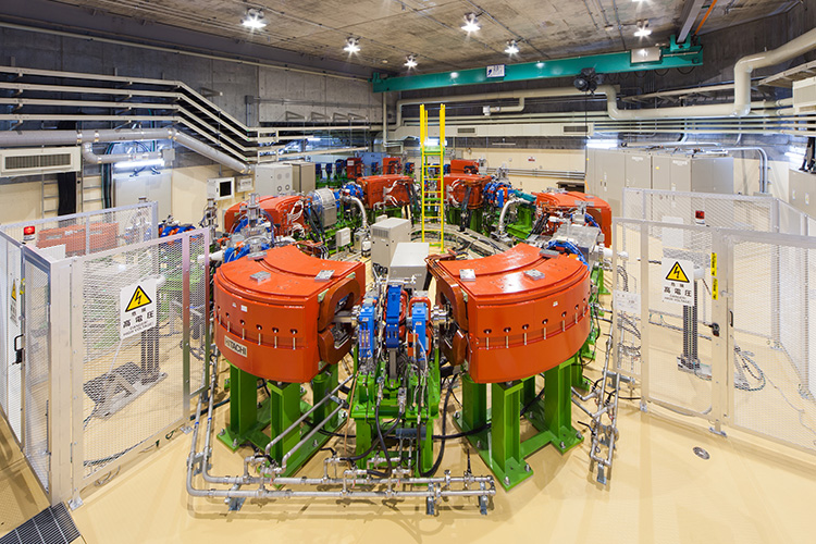
With a diameter of 7 m, and a 23 m-long circular path, this ring-shaped device takes in protons accelerated in the linac and increases their speed to 60% that of light. The synchrotron contains a vacuum pipe to transport the protons, electromagnets to curve their path, a high-frequency accelerating cavity, and other devices, each of which is accurately controlled to accelerate the protons. When the protons are accelerated, the accelerating cavity and electromagnets are controlled in sync with the protons, which is what gives the synchrotron its name.
The protons are flung straight ahead into the vacuum pipe, and curve due to the Lorentz force acting on them in the magnetic field created by the electromagnets. The direction that the protons curve follows Fleming's left-hand rule. This way, we can circulate the proton beam inside the beam pipe, following the ring-shaped track.
A synchrotron has a high-frequency accelerating cavity at a single location along the ring, which uses high-frequency electrical fields to accelerate the protons. In this high-frequency accelerating cavity, the electrical field is controlled to match the timing at which the protons pass through, thus accelerating them.
Using this device, we can circulate the protons inside the synchrotron anything from 100,000 times to a million times, reaching a maximum of 60% of the speed of light. All this takes under a second. The final speed we accelerate the protons too depends on the depth of the cancer, and is calculated so as to have the protons stop precisely at the tumor location.
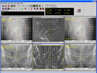
Treatment Planner
This device is used to plan the treatment, based on the data from the CT, MRI, and PET scans, etc., that were taken to plan treatment. The location and shape of the cancer for treatment are entered, and the direction and amount of protons to use are set, allowing us to assess and study the degree to which the radiation will hit the cancer and the surrounding healthy tissue. We treat people every day using this treatment planner.
CT Simulator
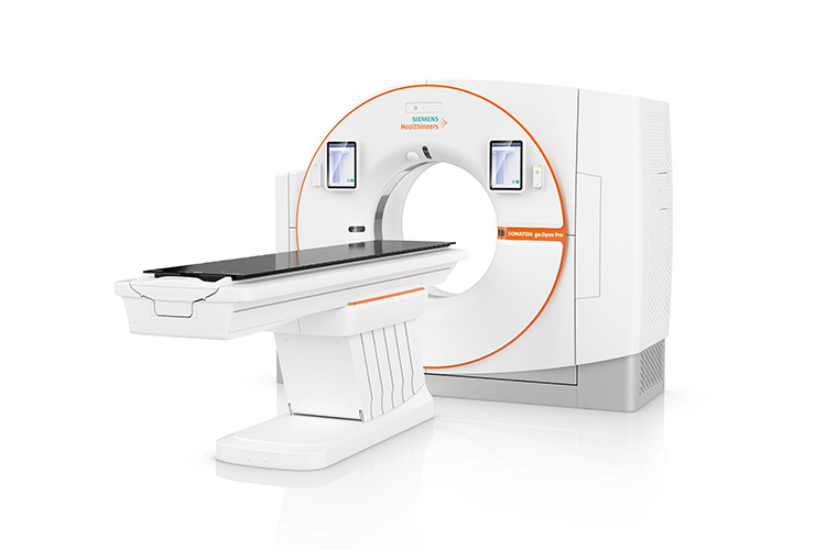
This takes the CT scans to use in treatment planning. The patient undergoes a CT scan in the same posture as will be used for the proton therapy. The opening is larger than a normal CT scanner, which means that there is less feeling of being enclosed, and it also lets us scan patients in the same posture they will use in their therapy without their elbows or other parts getting in the way. It uses a 16-row multi-slice CT system, which means CT scans can be taken that show breathing movement (a normal CT scan has three axes; this is called a four-dimensional CT scan as it adds the axis of time).